

|
Overview:
Rolls and colleagues discovered face-selective neurons in the inferior
temporal visual cortex; showed how these and object-selective
neurons have translation, size, contrast, spatial feequency and
in some cases even view invariance; and showed how neurons encode
information using mainly sparse distributed firing rate encoding. These
neurophysiological investigations are complemented by one of the few
biologically plausible models of how face and object recognition are
implemented in the brain, VisNet. These discoveries are complemented by
investigation of the visual processing streams in the human brain using
effective connectivity. Key descriptions are in 639, B16, 656, 682 and 508.
The discovery of face-selective
neurons (in the amygdala (38, 91, 97), inferior temporal visual cortex (38A,
73,
91,
96, 162), and orbitofrontal cortex (397))
(see 412, 451, 501, B11, B12, B16).
Our discovery of face selective neurons in the inferior temporal visual
cortex was published in 1979 (Perrett, Rolls and Caan 38A), and in the amygdala was published in 1979 (Sanghera, Rolls and Roper-Hall 38), with follow-up papers in 1982 (Perrett, Rolls and Caan, 73), 1984 (Rolls 91), and for the amygdala in 1985 (Leonard, Rolls, Wilson and Baylis 97).
These discoveries were followed up and confirmed (Desimone, Albright,
Gross and Bruce 1984 J Neuroscience). The discovery of face neurons is
described by Rolls (2011 501).
The discovery of face
expression selective neurons in the cortex in the superior temporal
sulcus (114,
126) and orbitofrontal cortex (397). Reduced connectivity in this system has been identified in autism (541, 609).
The discovery that visual
neurons in the inferior temporal visual cortex implement translation, view, size, lighting, and spatial frequency
invariant representations of faces and objects (91, 108, 127, 191, 248, B12, B16).
The effective connectivity of
the human prefrontal cortex using the HCP-MMP human brain atlas has
identified different systems involved in visual working memory (660).
In
natural scenes, the receptive fields of inferior temporal cortex
neurons shrink
to approximately the size of objects, revealing a mechanism that
simplifies
object recognition (320, 516, B12, B16).
Top-down
attentional control of visual processing by inferior temporal cortex
neurons in
complex natural scenes (445).
The discovery that in
natural scenes, inferior temporal visual cortex neurons encode
information
about the locations of objects relative to the fovea, thus encoding
information
useful in scene representations (395, 455, 516).
The discovery that the inferior
temporal visual cortex encodes information about the
identity of objects, but not about their reward value, as shown by reversal and devaluation investigations (32, 320, B11). This provides a foundation for a key principle in primates
including humans that the reward value and emotional valence of visual stimuli are
represented in the orbitofrontal cortex as shown by one-trial reversal learning and devaluation investigations (79, 212, 216) (and to some extent in the amygdala 38, 383, B11), whereas before
that in visual cortical areas, the representations are about objects and stimuli independently
of value (B11, B13, B14, B16). This provides for the separation of emotion from perception (B11, B16, 674).
The discovery that information
is encoded using a sparse distributed graded representation with
independent
information encoded by neurons (at least up to tens) (172, 196, 204, 225, 227,
321, 255, 419, 474,
508, 553, 561, B12, B16). (These
discoveries argue against ‘grandmother cells’.) The representation is
decodable
by neuronally plausible dot product decoding, and is thus suitable for
associative computations performed in the brain (231, B12).
Quantitatively
relatively little information is encoded and transmitted by
stimulus-dependent
('noise') cross-correlations between neurons (265, 329, 348, 351, 369, 517). Much of the
information is available from the firing rates very rapidly, in 20-50
ms (193,
197, 257, 407). All these discoveries are important in our
understanding of
computation and information transmission in the brain (B12, B16).
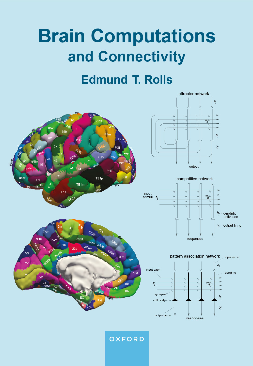
A
biologically plausible theory and model of invariant visual object recognition in the ventral
visual
system closely related to empirical discoveries (162,
179,
192,
226,
245,
275,
277,
280,
283,
290,
304, 312,
396,
406,
414,
446,
455, 473,
485, 516, 535,
536, 554, B12, 589, 639, B16).
This approach is unsupervised, uses slow learning to capture
invariances using the statistics of the natural environment, uses only
local synaptic learning rules, and is therefore biologically
plausible in contrast to deep learning approaches with which it is compared (639, B16).
A theory
and model of coordinate transforms in the dorsal visual system using a
combination of gain modulation and slow or trace rule competitive
learning. The theory starts with retinal position inputs gain modulated
by eye position to produce a head centred representation, followed by
gain modulation by head direction, followed by gain modulation by
place, to produce an allocentric representation in spatial view
coordinates useful for the idiothetic update of hippocampal spatial
view cells (612).
These coordinate transforms are used for self-motion update in the
theory of navigation using hippocampal spatial view cells (633, 662, B16).
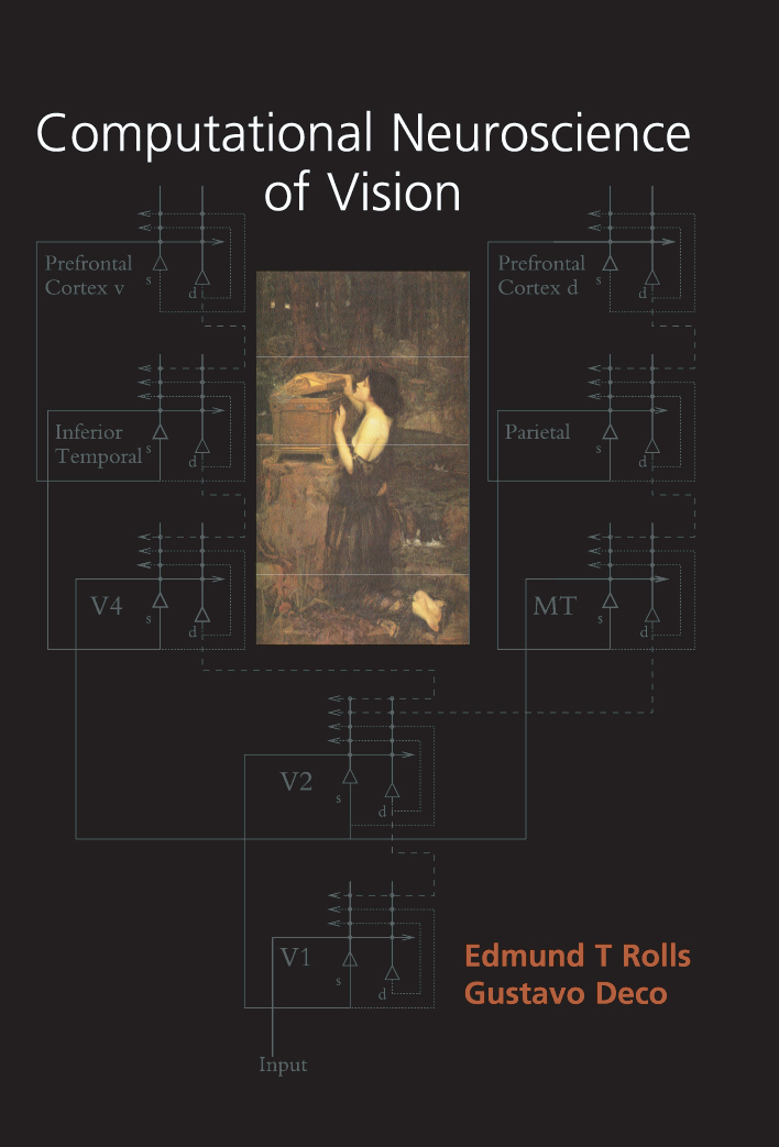
The
effective connectivity of the human visual cortical streams using the
HCP-MMP human brain atlas has identified different streams (656, B16, 682, 676).
A Ventrolateral Visual ‘What’ Stream for object and face recognition
projects hierarchically to the inferior temporal visual cortex which
projects to the orbitofrontal cortex for reward value and emotion, and
to the hippocampal memory system. A Ventromedial Visual ‘Where’ Stream
for scene representations connects to the parahippocampal gyrus and
hippocampus. This is a new conceptualization of 'Where' processing for
the hippocampal system, which revolutionizes our understanding of
hippocampal function in primates including humans in episodic memory
and navigation (682, 662). A Dorsal Visual Stream connects via V2
and V3A to MT+ Complex regions (including MT and MST), which connect to
intraparietal regions (including LIP, VIP and MIP) involved in visual
motion and actions in space. It performs coordinate transforms for
idiothetic update of Ventromedial Stream scene representations (682, 662). An
Inferior bank STS (superior temporal sulcus) cortex Semantic Stream receives
from the Ventrolateral Visual Stream, from visual inferior parietal
PGi (655), and from the ventromedial-prefrontal cortex reward system (649) and connects to
language systems (682, 654). A Superior bank STS cortex Semantic Stream receives visual
inputs from the Inferior STS Visual Stream, PGi, and STV, and auditory
inputs from A5, is activated by face expression, motion and
vocalization, and is important in social behaviour, and connects to
language systems (656, B16, 682, 654).
Binaural
sound recording to allow 3-dimensional sound localization (11A,
UK provisional
patent, Binaural sound recording, B16).
|